1.Introduction
Telemedicine, electronic patient records, online medical textbooks, remote education and so on just began to be put to practical use. Then a large amount of digitized images with input and output equipment for them are going to prevail among wide-ranging scenes of medical practice, but they have not well proved to be used for reliable medical diagnoses as usual analog images. This paper will present the present status of the research in this field, and the possible role of multispectral imaging in it.
2.Background
The purpose to use image data in medicine is to make an exact medical diagnosis. So our final target is to ensure that physicians can make the same diagnosis looking at the digital images as they are looking at real patients or long-established analog images.
The process of medical diagnosis based on usual visual data is broken down into several steps.
(A1) The subject of observation emanate proper light waves of various wavelengths. This steps is affected by the characteristics of the surface of that subject and the lighting. There may be penetrated light or invisible electromagnetic waves converted into visible light, etc.
(A2) The light waves pass through an iris, a lens and a vitreous body, and reach to the retina. This steps is affected by the color of the iris and the transparency of the path of the light waves.
(A3) The rod cells convert the brightness of the light waves into frequency modulated pulse signals and the three kinds of cone cells convert the color of the light waves into pulse signals, and they are transmitted to optic nerves. There may be many interactions between cells, but most processes are unclear. One important fact is that adaptation of the human retina to surrounding lighting is greatly affected to the signals produced by the sensor cells in the aspects of both color and brightness.
(A4) The signals are transmitted to the brain and converted into image data. This step is mentioned as visual sensation and includes the specialized process of various image abstraction.
(A5) The image is connected to some of the concept accumulated in the memory and given particular meanings. This step is mentioned as visual recognition and the most of its process is unclear.
(A6) The concept is compared to the experiences accumulated in the brain, and deducted by the knowledge established in the brain. This step is mentioned as decision making and its process is in the mystery yet.
On the other hand, the process of medical diagnosis based on digital images are as follow:
(B1) Same as (A1).
(B2) The light waves are converted into digital image data which are composed of color information of each pixel represented by RGB system and the coordinates of the pixel on the two-dimensional plane. In this step, not only a large part of information that original lights have is lost, but also various biases are mixed which depend on the characteristics of equipment used for A-D conversion.
(B3) The digital image data is stored or transferred to be used at another time or place.
(B4) According to the color information of each pixel of the digital image data, the R, G and B dots of the display equipment corresponding to the pixel emanate red, green and blue light each with appropriate combination of intensities to give the same stimulus to retina as the light wave which has that color. Because there is a large discrepancy between the wavelengths of red, green and blue lights and that of the peaks of the response curve of three types of the cone cells, some kinds of approximation are indispensable in this step.
(B5) The light waves pass through an iris, a lens and a vitreous body, and reach to the retina. This steps is affected by the color of the iris and the transparency of the path of the light waves. Unlike natural lights, these lights include only three bands of the wave length. Therefore, these influences may cause different results from (A2).
(B6~9) Same as (A3~6)
The steps from (B2) to (B5) are possibly cause to wrong diagnoses.
3.Recent Progress of the Research [1]
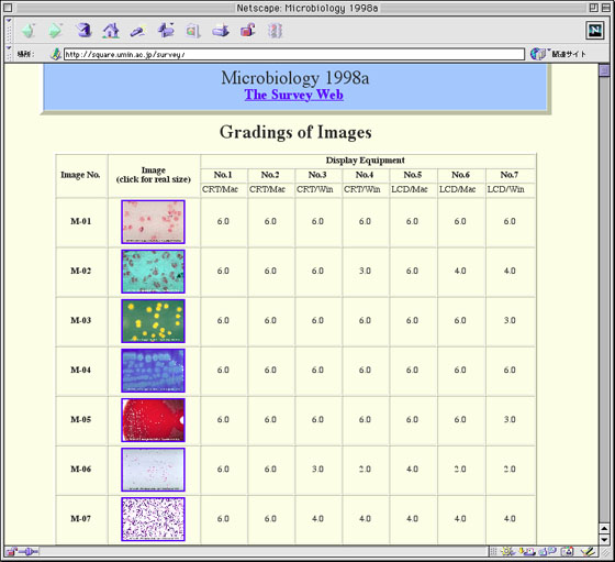
One of the earliest investigation about the color of digital imaging in medicine is made by the Morphological Internet Survey Research Project Team. The Ministry of Science, Education and Culture of Japan organized it in 1998, and it consists of nine researchers from seven Universities and 30 co-researchers from various fields of laboratory medicine and clinical pathology in Japan (in detail, consult its home page at
http://square.umin.ac.jp/survey/).
The following are overview of the research.
(1) Pictures of typical specimens from urinary tests, hematology, microbiology, immunology, physiology and pathology were digitized and the diagnostic reliability were evaluated using various display equipment including a extra high resolution (QSXGA, 200 pixel per inch) LCD.
(2) The qualities of most of digitized pictures properly prepared are almost the same as slidefilms, but some of them required to be observed with the QSXGA display to maintain required qualities.
(3) Besides, there are large variations in reproduction of colors among the displays, which may incidentally cause erroneous diagnoses (Fig. 1).
(4) To overcome this problem, a new calibration system based on a novel way of thinking has been developed.
(5) To discuss the same problem possibly arising in other medical fields, the 1st Symposium of the 'Color' of Digital Imaging in Medicine was held on 8th-9th May, 1999 at Tokyo Medical and Dental University, and understanding about this problem and a consensus of the orientation toward its solution were well advanced (in detail, consult a home page at
http://square.umin.ac.jp/medicolor/)[2].